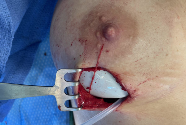
Take a look at the first picture- it is of a midline incision in the back of the neck. Can you identify the nerve by the black arrow and the nerve by the blue arrow? Spoiler alert……don’t look at the second picture just yet.
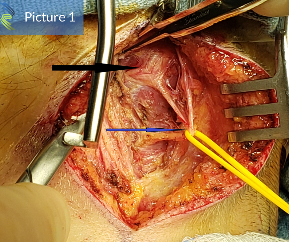
In the second picture, can you identify the same two nerves clearly seen communicating. How about now….which nerves are these? Don’t look at the third picture yet.
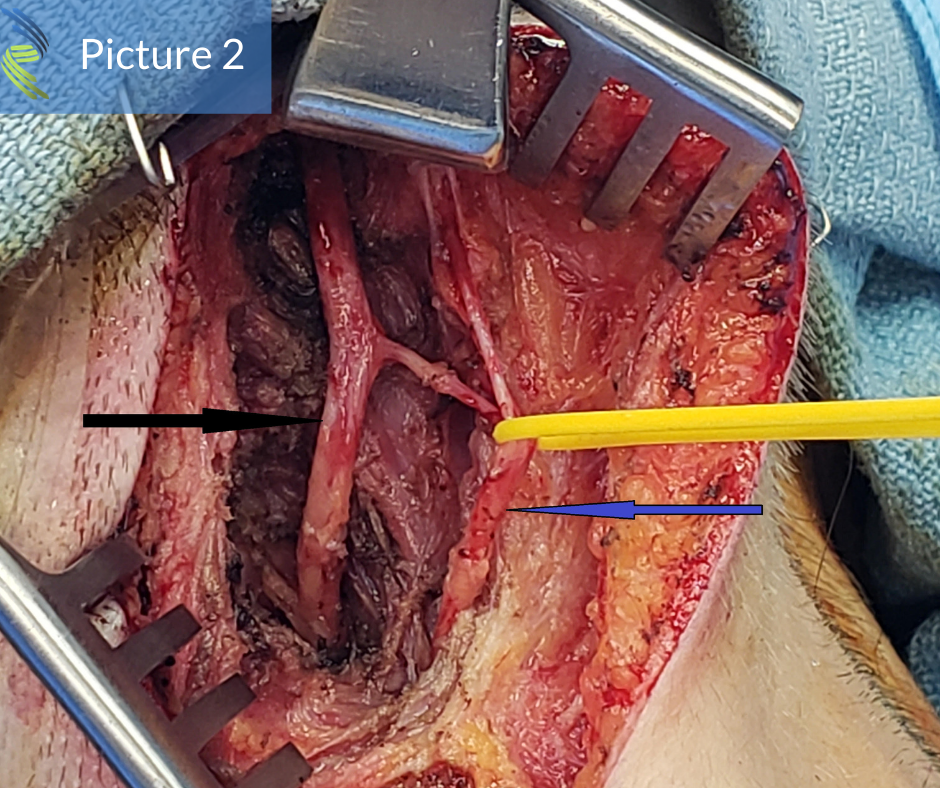
Now in the third image, the greater occipital (black arrow in the previous images) and third occipital (blue arrow in the previous images) nerves are clearly marked and are separate.
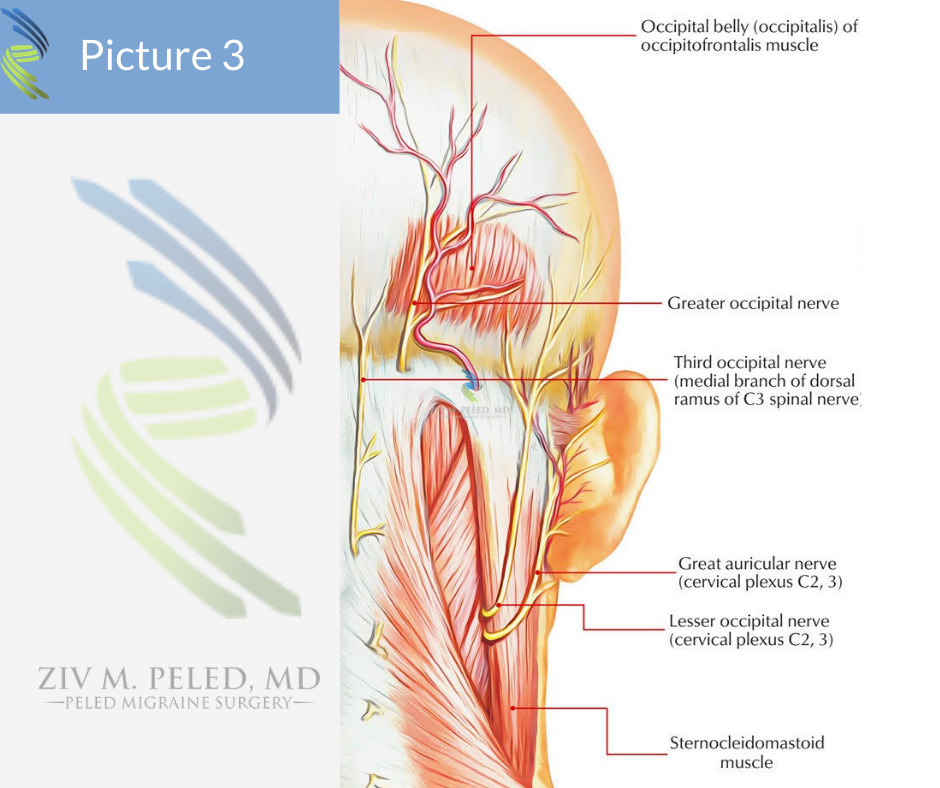
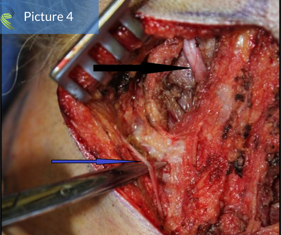
In the fourth image, the same two nerves are seen with the same delineating arrows in a more classic relationship. However, notice that even in this last picture, the third occipital nerve is lateral to the greater occipital nerve which is also slightly different than in the illustration.
The take home message with this post is that anatomy can be variable or as I tell my patients, “Everyone is wired differently.” Appreciation of this fact is one of the many reasons experience counts when choosing a headache surgeon.