
Ziv M. Peled, MD is a Plastic & Peripheral Nerve Surgeon specializing in chronic headache/migraine relief. Board-certified and Harvard-Trained, he is committed to raising awareness and educating others about the life-changing benefits of headache/migraine surgery. While offering exceptional and compassionate care, Dr. Peled’s mission is to bring positive and substantive change to his patients’ lives.

UNDERSTANDING
YOUR MIGRAINE
UNDERSTANDING
YOUR MIGRAINE
OCCIPITAL
FRONTAL

TEMPORAL
OCCIPITAL
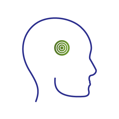
The occipital region is the area in the back of the scalp/head. It is one of the most commonly treated areas for patients seeking surgical relief from chronic headaches. In my humble opinion, many people are often told they suffer from migraines, when they really have neuralgia (i.e. pain caused by mechanical compression or irritation of a nerve). The offending structure(s) causing this compression/irritation may be a spastic muscle, an enlarged or aberrant blood vessel, and/or tight connective tissue. When the pain emanates from and/or occurs in the occipital region of the head, it is called ‘occipital neuralgia’ or ON for short. In the case of people who have pain in the back of the head, the question for the surgeon is, which “neur” (i.e. nerve) is causing the “algia” (i.e. pain).
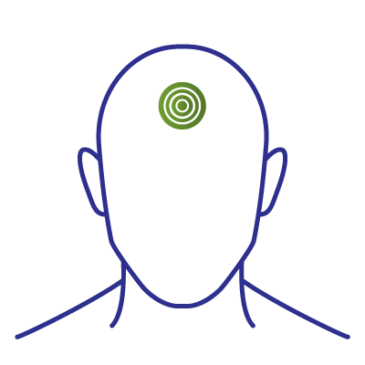
FRONTAL
The frontal region is located, as the name suggests, in the front of the head, specifically the forehead and glabella (that region between your eyebrows). When this type of headache pain occurs secondary to nerve compression and/or irritation, it is often secondary to pathology within the supraorbital and/or supratrochlear nerves which are branches of the trigeminal nerve. In some cases, an anterior branch of the zygomaticotemporal nerve (see temporal link) may travel toward the side of the forehead and contribute to the overall, frontal headache complex. It used to be thought that the supraorbital and supratrochlear nerves were primarily compressed by the glabellar musculature, specifically the corrugator supercilii, procerus, and depressor supercilii muscles. However, more recently it has been demonstrated that compression of both nerves may also occur at the orbital rim (i.e. the top of the eye socket) as these nerves sometimes emerge from behind and above the eye through very tight fascial openings or through bony foramina as opposed to notches within the orbital rim. In these locations, adjacent vessels can also act as compressive structures.
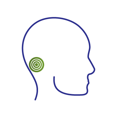
TEMPORAL
The temporal region is located at the side of the head/scalp – i.e. the temples. Most commonly, headache symptoms in these areas secondary to peripheral nerve irritation/compression are a result of 2 possible, offending nerves. One is called the auriculotemporal nerve and the other is called the zygomaticotemporal nerve and both are branches of the trigeminal nerve. The former is located just above the helical root which is where the external ear (i.e. pinna) meets the scalp and often sends branches ascending toward the vertex of the skull as well as in front of and occasionally behind the ear. In this area, the auriculotemporal nerve may be compressed by the adjacent connective tissue and superficial temporal artery and/or vein which may be enlarged, in an aberrant location, or even wrapping around the nerve itself like an anaconda. The zygomaticotemporal nerve is located just to the side of the eye socket toward the front of the temple. If you clench your back teeth, you might feel a bulge in this area as the temporalis muscle contracts. In this location, the zygomaticotemporal nerve may be entrapped by connective tissue overlying the temporalis muscle which is too tight and/or an adjacent blood vessel. In addition, the zygomaticotemporal nerve may have “downstream” branches that, like the auriculotemporal nerve, go towards the top of the scalp, anteriorly towards the forehead or posteriorly toward the ear. There can be an overlap in the sensory distribution of these two nerves in the mid-portion of the temple which can make it difficult to discern which one of these nerves or both nerves are involved in temporal pain generation.
OUR APPROACH
When traditional methods have failed, we are often able to help. Many chronic headache/migraine patients actually suffer from nerve compression that is similar to that which I have been treating for many years in other parts of the body. Our approach focuses on determining whether there are any peripheral nerves in the head or neck area that could account for chronic headache/migraine symptoms using a combination of a thorough history, comprehensive physical exam and diagnostic nerve blocks.
My results have been very positive and have validated my decision to make helping chronic headache/migraine sufferers a primary career goal. It is estimated that there are more than 39 million people diagnosed with migraines in the United States alone. Furthermore, more than 6.5 million Americans (and many more people globally) are afflicted by chronic migraines, those occurring more than 15 days/month! It has been and continues to be my privilege to make a difference in these patients’ lives.
SCHEDULE A CONSULTATION
We provide a range of emergency, medical, general, residential, and community health services.